definition of articulation
Classification of joints according to compositional organization and mobility
Joints are classified into fibrous joints, cartilaginous joints, and synovial joints according to the structure of the joint. It is also classified according to the range of motion, but the wider the range of motion, the more flexible the organization is. Therefore, starting an approach from one (organization or mobility) perspective naturally converges to the opposite (mobility or organization) criterion.
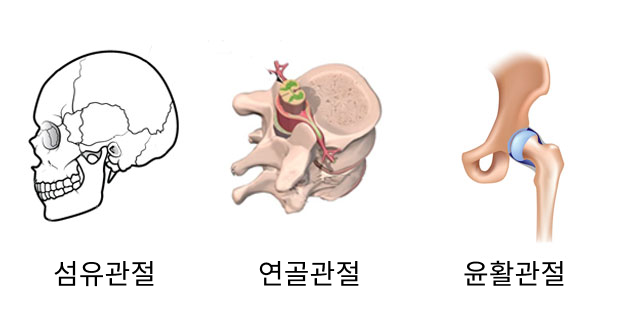
The bones in contact are connected by fibrous connective tissue, most often in the case of immobilized joints (skull, teeth, ribs, etc.).
Fibrous joints are further divided into suture joints, ligamentous joints, and formal joints.
Suture joint: Cranial joint with no mobility (skull)
· Ligamentous Joint: Two bones are joined by an interosseous ligament (radioulnar joint)
Canonical joint: A form in which the end of one bone enters the hole of the bone where it touches (teeth)
The joint between the two bones is joined by cartilage, so a 'semi-movable joint' with a narrow range of motion is a cartilaginous joint. spine, etc.
Cartilage junction: joint between rib and breastplate
Fibrochondral junction: additional disc, pubic symphysis, sternum disease cartilage joint
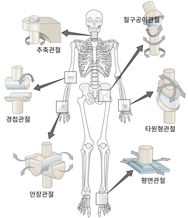
In the most common type of joint, the space between two bones is covered with articular cartilage. Between these articular cartilage is a joint capsule (joint capsule) filled with synovial fluid secreted by the synovial membrane, so it is also called a synovial joint. It enables relatively free movement, and each epiphysis faces each other.
Articular space: space filled with synovial fluid at the point where the adjacent bones are located
Articular Cartilage: Cartilage covering the articular surfaces
Joint capsule: the strongest fibrous connective tissue. wraps around the ends of two bones

Classification according to the type of joint connection
Joints are also classified according to the shape in which two bones meet or the axis of movement.
One bone rotates within a link formed by another bone. Located at the top of the neck between the atlas and cervical vertebrae, it enables left and right rotation of the head.
It is a type of joint in which the end of a spherical bone is inserted into the empty space at the end of another bone.
It is formed by saddle-shaped bones that meet vertically. It has a narrow range of motion and can mainly move left and right or back and forth. The lower thumb joint is the only saddle joint.
It is a shape in which an oval cup-shaped rice tip and an oval cup-shaped bone tip are fitted. It moves in most directions, but rotation may be limited. A typical elliptical joint is the wrist.
Flat surfaces slide back and forth or side to side. There are foot and wrist joints, etc.
The circumferential surface of the bone is inserted into the groove of the bone in contact with it, enabling the motion of bending and extending the limb. The knee, elbow, and finger joints are hinge joints.

Joint disease prevention and opportunities
Joint disease is caused by arthritis, injury or infection, aging or disease. Except for acute pain such as accidents, it is difficult to detect until the pain occurs, and because the degree is gradual, patients sometimes become insensitive to the intensity of the pain. Also, according to the principle of the kinetic chain (Kinetci Chain), diseases that occur in one place may affect other parts.
In the end, when you visit a hospital, it takes a lot of time to normalize or it is impossible to treat.
Therefore, hospitals will be able to save opportunities for positive experiences if they more aggressively suggest precursor symptoms and examination methods for musculoskeletal pain, including joint disease, to patients waiting for treatment in the hospital lobby.
More News & Media